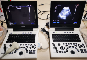
TH-300C LAPTOP Color Doppler System
- Home
- service
- RADIOLOGY EQUIPMENT
- TH-300C LAPTOP Color Doppler System
GET A QUOTE
OVERVIEW
What you need to know
An Excellent Entry Level laptop Color Doppler Machine:
TH-300C offers decent 2D images with THI, excellent CFM images, as well as Doppler imaging modes such as CFM, PW, PDI, etc.
Features & Advantages:
- Multi-application: abdomen, OB/GYN, small parts, peripheral vessels, urology, cardiac, Rectum, pediatric, orthopedic, galactophore, intra-operative, ultrasound guided biopsy.
- Continuous high-precision DBF – High Resolution Imaging
- Parallel scanning – Faster 2D frame rate.
- Parallel scanning – Faster 2D frame rate.
- PC platform / Windows 7 Embedded O/S
- Simple, direct and user-friendly workflow
- Customized PC motherboard ensures system stability and reliability
- Write protection of ultrasound control program, safe from virus attack and power outage
- Operation system restoration by one click
- Complete Digital Solution:
- Digital core module with DBF
- Digital display with DVI, enabling enhanced screen display
- Digital printing, enabling clearer printing with no need of adjustment
- Transducers:
- Self-owed transducer workshop
- Multi-frequency transducer series
- Max frequency up to 12MHz
- Multi-language: English, French, Spanish, etc
- Printing:
- Color printers supported
- All PC and Video Printers supported
- Direct printing by one click
Standard Configuration:
TH-300 main unit:
- 12″ high – definition LED monitor
- Two activated transducer connectors
- Min. 300-frames of Cine Loop
- B / B+B / 4B / B+M / B-steer (note) / Pulsed Wave Doppler (PWD) / Auto-IMT
- Tissue Harmonic Imaging (THI) / SRI (Speckle Reduction Imaging)
- One Click Restoration of O/S
- 2 USB ports & one HDMI & one Audio out & VGA & DICOM 3.0
- Panoramic zoom
- Triplex
- Measurement & calculation software package
- Electronic convex array transducer: CA3.5MHz/R60 (2.0-6.0MHz)
- Li-ion Battery
- Auto – IMT
Technical Specifications:
General Descriptions
- Imaging mode: B, B+B, 4B, B+M, M,B/PW
- Gray scale: 256
- Display: 15″ high-resolution non-interlaced monitor, special for medical imaging
- Transducer frequency: 2.0 ~ 10MHz
- Transducer Connector: 2
- Image Technology: Continuous High-precision Digital Beam-former
- Dynamic Frequency Integration Imaging
- High-precision Dynamic Receiving Focus
- Super Wide-band Imaging Technology
- Self-adaptive Image Optimization Processing
- Multi-beam Imaging
- Self-adaptive Vascular Imaging
- Self-adaptive Doppler Imaging
- THI (Tissue Harmonic Imaging)
- Export function: Archiving to DVD (optional), DICOM, USB drive.
- Format: BMP, JPG, AVI, MP4
Measurement & Calculation:
- B-mode: distance, circumference, area, volume, angle, residual urine volume, histogram, profile
- M-mode: distance, time, velocity, heart rate
- D-mode: Doppler blood flow measurement, velocity, acceleration, pressure gradient, time, VI, PI, RI, etc
Software packages:
- GYN: uterus, endometrium, ovary, cervix, ovarian follicle
- OB: GS, CRL, LV, BPD, OFD, HC, TAD, LVW, HW, TCD, IOD, OOD, BD, APTD, TTD, AC, APD, FTA, HL, ULNA, FL, FIB, CLAV, etc
- Cardiac: M. Simpson, B-EF, M-EF (Pombo、Gibson、Teichholz), Diameter Function, PV flow, AV-Area, B-LV/Ao、M-LV/Ao, MV Regurg, etc
- Urology: volume of prostate, volume of bladder, volume of urine, volume of trans zone, Hip J.Angle(hip joint dislocation in neo-natal and babies), Slice V, etc
- Small Parts and Peripheral Vessels
- vascular cross-sectional area, heart rate, stroke volume, flow per unit time, Ejection Time, % stenosis, mean velocity of flow, RI, PI, etc.